X-Rays in Medical Diagnosis
The nature of X-rays and the range of frequencies used
X-rays are a form of ionising electromagnetic radiation and have a very high frequency and a very short
wavelength.
Their wavelengths range between 0.001 to 10 nm.
The technology used in X-ray production
 [Image credit: Jacaranda Physics 1 2nd Edition © John Wiley & Sons, Inc.]
An X-ray tube works as follows:
- The heated filament is positively charged and the tungsten target is negative.
-
Electrons are emitted from the heated filament towards the tungsten target due to the very high
potential difference between them.
- The tungsten target absorbs the electrons and releases some of the energy in the form of X-rays.
This process is very inefficient however and a lot of energy is released in heat.
For this reason the tungsten target has a copper mounting because it conducts heat and is cooled with by
circulating oil through the mount.
Spinning the tungsten target at high speed also helps to stop it overheating.
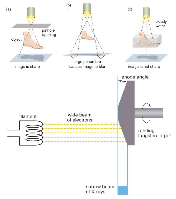 [Image credit: Jacaranda Physics 1 2nd Edition © John Wiley & Sons, Inc.]
Narrower beams of X-rays will produce a sharper image.
The tungsten target is therefore angled so that a wide beam of electrons will produce a narrow beam of
X-rays.
The clinical application of X-rays to form images
Hard and soft X-rays
Hard X-rays are X-rays with a higher frequency and are more penetrating than soft X-rays.
Soft X-rays are usually filtered when doing a scan because they can't penetrate through a patient's body
and add needless risk of radiation damage.
Attenuation
Attenuation is a measure of how much something absorbs X-rays.
The amount of attenuation increases with atomic density (number of protons in the nuclei).
For example,
bones have a higher attenuation than soft tissue and therefore bones produce a dark shadow when X-rayed
where as soft tissue appears much fainter.
Frequency
When a patient has an X-ray,
they are usually scanned at a frequency of approximately 7×10⁸ Hz because body tissues absorb this
frequency the best.
Use of X-rays in various parts of the body
X-rays are best suited to imaging bones and have a very high resolution.
For imaging soft tissue however, there is very little contrast and so a contrast medium is needed.
Contrast mediums are substances given to the patient that absorb X-rays and produce an image of the area
under investigation when X-rayed.
Usually CAT (Computer Axial Tomography) scans,
in which a series of X-rays are taken from various angles and interpreted by a computer,
are better for imaging soft tissue.
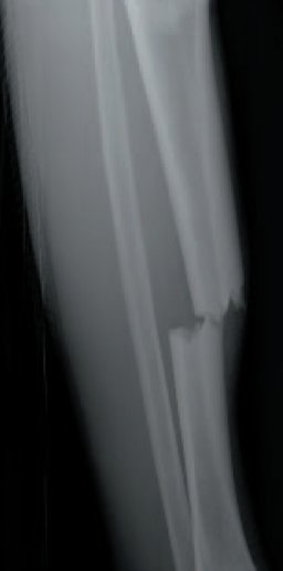
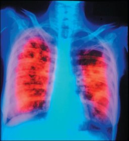
Examples of images attained using X-ray technology
This is an X-ray of a broken leg and shows that a bone has been broken.
|
 |
This X-ray shows that the lung on the right is damaged by tuberculosis.
|
 |
|