FAQ Sheet
- Using Radioactivity as a Medical Diagnostic Tool
Q. I'm trying to give a patient a thyroid investigation
and my colleague has suggested using radioisotopes.
What are the properties of radioisotopes that make them useful for medical diagnosis?
- Dr Tim (Doctor)
A. Radioisotopes are useful as diagnostic tools because:
-
A gamma camera can be used to detect how much of the isotope is metabolised in order to give an
indication of how healthy an organ is and to detect growth of tumours.
- They can be used to label compounds that can then and traced to show functional information.
- The use of radioisotopes for diagnosis is relatively safe.
Q. I'm working as an apprentice for a doctor and next
week I'll have a chance to image a patient using radioisotopes by myself. However, I don't know which
radioisotope to use. How do you know which is the correct one?
- Matt (Apprentice)
A. The half-life plays an important role in choosing the
correct isotope:
-
If the radioisotope remains in the patient's body for a long time after the scan,
the half life must be long enough for a scan to be taken, but short enough that it will be mostly
decayed shortly after the scan so that the patient does not remain radioactive.
-
If the radioisotope is metabolised and leaves the body shortly after the scan:
Radioisotopes with longer half-lives are suitable for use because they will leave the body and the
patient will no longer be radioactive.
The radioisotope chosen must also be able to label a compound to produce a suitable radiopharmaceutical
that will be metabolised by the area of interest.
For example, iodine is metabolised by the thyroid,
which makes iodine-123 useful for thyroid investigations.
Gamma-emitting sources are the best isotopes to use for nuclear medicine.
Q. In your last response you said that
"Gamma-emitting sources are the best isotopes".
Why?
- Matt (Apprentice)
A. Gamma radiation is the most useful for diagnosis
because gamma rays are highly penetrating and not as ionising as alpha or beta particles.
This means that they can be detected outside the body and will cause minimum damage to the patient.
Since gamma decay is due to the nucleons rearranging themselves,
the daughter nucleus of gamma decay is stable and the same isotope as the parent,
which makes it safer than alpha or beta decay that produces new isotopes when they decay.
Q. I'm a student currently studying medical physics at
school and I was wondering what radiopharmaceuticals were and how they're produced?
- Andrew (Student)
A. A radiopharmaceutical is a compound that has been
labelled with a radioisotope and can then be traced with a gamma camera.
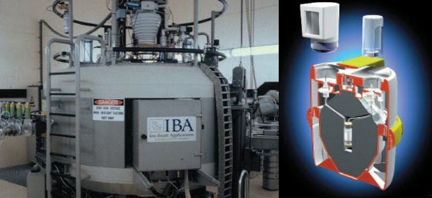
The radioisotopes can be produced in a nuclear reactor or a cyclotron and then delivered to the hospital or
milked from another isotope in a generator.
The radioisotope is then chemically attached to the compound to produce a suitable radiopharmaceutical that
will be metabolised by the area of interest.

Q. I'm still confused.
How is a radioisotope introduced to the site to be diagnosed?
- Andrew (Student)
A. As mentioned before,
the radioisotope is attached to a compound to produce a radiopharmaceutical that will be metabolised by the
area of interest.
The radiopharmaceutical is then injected, inhaled or drunk by the patient.
The radiopharmaceutical will travel through the body to the site to be diagnosed and the radioisotope will
then build up in the areas that metabolise the compound the most.
Q. I can never remember which radioisotope is the best
one for the job.
Could you please give me a list of all the different radioisotopes and their uses?
- Dr Tim (Doctor)
A. There are many different radioisotopes and ways that
they can be used, so only a few common ones are listed here:
-
Technetium-99m
Technetium-99m is used for imaging bones, the brain and the lungs.
When imaging the bones,
the Technetium-99m is injected into the body and it will build up around the areas of most blood flow.
Hot spots are where the blood flowed most and usually indicate a disease.
Similarly when imaging the brain,
the activity of various parts of the brain can be measured in order to diagnose patients for brain
damage.
In order to check whether airways are clear,
the patient inhales the technetium and an image is taken with a gamma camera.
Cold spots where there is no radiation indicates that the airway to that part of the lung is blocked.
-
Iodine-123
Iodine-123 is used for thyroid investigations because it is metabolised by the thyroid gland.
The patient drinks the iodine as a salt solution.
The iodine is then metabolised by the thyroid and an image can be taken with a gamma camera.
This can be used to check for goitre, which is due to an enlarged thyroid gland.
-
Thallium-201
Thallium-201 is used for imaging the heart.
It is injected into the patient and can then be traced.
An image produced with a gamma camera can be used in order to determine the position of damaged heart
muscles.
|
