Ultrasound in Medical Diagnosis
The wave properties of ultrasound
 [Image credit: Jacaranda Physics 1 2nd Edition © John Wiley & Sons, Inc.]
Ultrasound relies on high frequency sounds to image the body and diagnose patients.
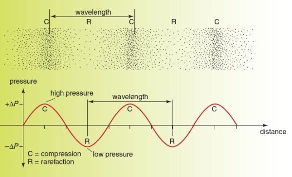
Ultrasounds are therefore longitudinal waves which cause particles to oscillate back and forth and produce
a series of compressions and rarefactions.
Using the following formula,
it is possible to calculate the velocity,
frequency or wavelength of a wave if the other two values are known:
v = fλ
Where:
- The velocity (v) is the speed of the wave. It is measured in m s-1.
- The frequency (f) is the number of times a particle oscillates per second. It is measured in Hz.
-
The wavelength (λ) is the distance between two compressions or rarefactions.
It is measured in m.
The amplitude is the distance a particle moves back or forth.
Compressions are areas of the wave where particles are close together and there is high pressure.
Rarefactions are areas of the wave where particles are far apart and there is low pressure.
The frequencies used in ultrasound diagnosis
Ultrasound uses high frequency sounds that are higher than the human ear can hear.
ie. 20 000 Hz.
Ultrasound can't detect objects that are smaller than its wavelength and therefore higher frequencies of
ultrasound produce better resolution.
On the other hand, higher frequencies of ultrasound have short wavelengths and are absorbed easily and
therefore are not as penetrating.
For this reason high frequencies are used for scanning areas of the body close to the surface and low
frequencies are used for areas that are deeper down in the body.
These frequencies generally range between 1-50 MHz.
How ultrasound is produced and detected
 [Image credit: Jacaranda Physics 1 2nd Edition © John Wiley & Sons, Inc.]
Ultrasound is produced and detected using an ultrasound transducer.
Ultrasound transducers are capable of sending an ultrasound and then the same transducer can detect the
sound and convert it to an electrical signal to be diagnosed.
To produce an ultrasound,
a piezoelectric crystal has an alternating current applied across it.
The piezoelectric crystal grows and shrinks depending on the voltage run through it.
Running an alternating current through it causes it to vibrate at a high speed and to produce an ultrasound.
This conversion of electrical energy to mechanical energy is known as the piezoelectric effect.
The sound then bounces back off the object under investigation.
The sound hits the piezoelectric crystal and then has the reverse effect
- causing the mechanical energy produced from the sound vibrating the crystal to be converted into
electrical energy.
By measuring the time between when the sound was sent and received,
the amplitude of the sound and the pitch of the sound, a computer can produce images,
calculate depths and calculate speeds.
The nature of A-scans, B-scans, sector scans and phase scans
A-scans
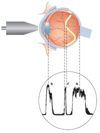
A-scans can be used in order to measure distances.
A transducer emits an ultrasonic pulse and the time taken for the pulse to bounce off an object and
come back is graphed in order to determine how far away the object is.
A-scans only give one-dimensional information and therefore are not useful for imaging.
|
 |
B-Scans
B-scans can be used to take an image of a cross-section through the body.
The transducer is swept across the area and the time taken for pulses to return is used to determine
distances,
which are plotted as a series of dots on the image.
B-Scans will give two-dimensional information about the cross-section.
|
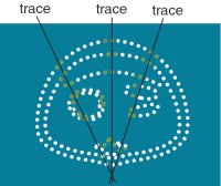 |
Sector scans
Sector scans can be used to take a sector shaped image of the body.
The transducer is swept back and forth across the area,
producing a series of B-scans that build up an image.
Sector scans are much harder to produce than phase scans and are much less common.
However,
they are useful for taking images when there is only a small amount of space.
|
 |
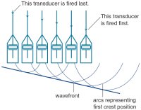
Phase scans
Phase scans have hundreds of transducers in the same probe in order to take a high resolution
real-time scan.
The angle of the wavefront can be altered by firing the transducers one after another,
when this happens they are out of phase.
By changing the angle of the wavefront,
a three-dimensional image can be built up over a large area.
|
 |
[Image credit: Jacaranda Physics 1 2nd Edition © John Wiley & Sons, Inc.]
Examples of images attained using ultrasound technology
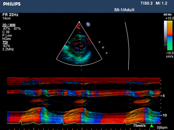
[Image credit: Philips Medical/Ultrasound & Monitoring Systems © Philips]
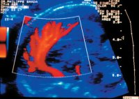
This image is of some tissue and demonstrates how ultrasound can image blood flow using the Doppler
effect.
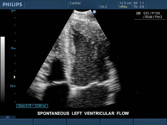
[Image credit: Philips Medical/Ultrasound & Monitoring Systems © Philips]
This is an image obtained of the heart's left ventricle.
|