Medical uses of Endoscopes and Lasers
The operation of optical fibres
Optical fibres are narrow tubes of glass fibres with a plastic coating that carry light from one end to the
other.
The light bounces off the walls of the fibre and can even bounce around corners.
The properties of optical fibres make them useful for a wide range of applications including:
- Medical - to transmit pictures of organs and arteries
- Industrial - to transmit pictures of the inside of complex machinery
- Communications - to transmit data over long distances without transmission loss
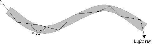
Light rays use total internal reflection to travel along the fibres.
In order for this to be achieved, the light ray must hit the walls of the fibre at a minimum angle of
82°, which is the critical angle for light travelling from glass to plastic.
Since the fibres are very narrow, this is usually not a problem.
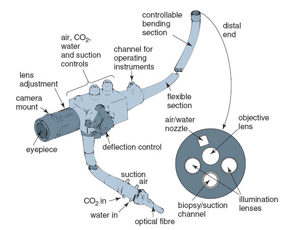
The parts of an endoscope
 [Image credit: Jacaranda Physics 1 2nd Edition © John Wiley & Sons, Inc.]
Shaft
The shaft is only 10mm in diameter and can be up to 2 metres long.
It is flexible and coated in steel and plastic in order to make it waterproof,
prevent chemical damage and to make it easy to manoeuvre through the body.
It has contains:
-
Fibre optic bundles
Light is guided to the area under investigation by non-coherent fibre optic bundles
(bundles where the optical fibres are not lined up at both ends).
However, the image must be transmitted back by a coherent fibre optic bundle
(a bundle where the optical fibres are lined up at both ends of the fibre so that an image can be
transmitted).
In order to produce a clear image, the shaft contains up to 10 000 fibres!
-
Water Pipes
Carry water to wash the lens and keep the view clear.
-
Operations channel
Carries accessories to the distil end for surgery.
-
Control cables
Controls which way the distil end is bent.
-
Aditional optional channel
Carries air or carbon dioxide to and from the distil end
Distil End
The distil end is inserted into the patient's body.
There are controls on the viewing end to make it bend in the desired direction.
The image is focused by a lens on the end.
Uses of endoscopes
Five medical procedures carried out using an endoscope:
-
Arthroscopy
The endoscope is inserted through an incision in the skin near a joint under investigation.
This can be used to look at the joint and preform operations such as removing torn tissues.
-
Bronchoscopy
The endoscope is inserted through bronchial tubes within the lungs in order to look at the airway and to
remove any objects blocking the airway.
-
Endoscope Biopsy
The endoscope is inserted through an incision or opening in the body that leads to the area under
investigation.
Biopsy forceps are then used to take a sample of tissue that can then be analysed by a pathologist.
-
Gastroscopy (Also called Oesophagogastroduodenoscopy)
The endoscope is inserted down the throat to look for problems with the oesophagus,
stomach and duodenum such as bleeding or ulcers.
-
Laparoscopy
The endoscope is inserted through an incision in the abdominal in order to look at abdominal organs and
preform minor surgery.
Advantages of using endoscopes
The use of endoscopes is much less invasive than open surgery because only a small incision in the body is
required where as open surgery requires deep incisions.
This also means that recovery is quicker and there is less swelling, scarring and risk of infection.
Endoscopes can be used by an outpatients department and does not need to be done by a hospital.
This reduces costs.
Keyhole surgery
Keyhole surgery involves the use of lasers with endoscopes.
Lasers are useful for such surgery because:
-
They can shine high intensity light down an endoscope that can be focussed for cutting or destroying
tissue.
- They produce heat that causes the tissue around the cut to seal and prevents bleeding.
- The beam is narrow and can therefore make very precise and accurate cuts.
- Different frequency lasers can be used depending on the area being targeted.
Other medical uses for lasers
Due to their high intensity, narrow beam and high precision,
lasers can be used for surgery that was previously extremely difficult and dangerous.
They are now used in cosmetic surgery for removal of scars, wrinkles, birthmarks,
blood vessels and hair and can also be used in laser eye surgery to alter the surface of the cornea on a
microscopic scale in order to correct sight.
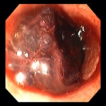
Images of internal organs obtained using an endoscope
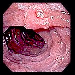
This image was taken of a patient with hematemesis.
This image revealed that it was due to a bleeding ulcer in the oesophagus.
|
 |
This image shows an elastic hair tie that was swallowed by a patient.
|
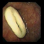 |
This is an image of a sessile polyp in a patient's duodenum.
By using biopsy,
it was found that it was a tubulovillous adenoma.
|
 |
[Image credit: The Atlas of Gastrointestinal Endoscopy © Atlanta South Gastroenterology, P.C.]
|